Reflectance Confocal Microscopy

Reflectance Confocal Microscopy: Seeing Cellular Dimensions

Reflectance Confocal Microscopy (RCM) is a non-invasive imaging technique that uses focused light to capture high-resolution, in vivo images of skin layers. It provides detailed visualization of cellular structures, essential for dermatology, skin cancer diagnosis, and real-time medical assessments.

Reflectance Confocal Microscopy

Overview

Reflectance Confocal Microscopy (RCM) stands at the forefront of cutting-edge imaging technologies, offering scientists and researchers an unprecedented glimpse into the microscopic realm of living tissues. This non-invasive imaging technique has revolutionized the field of dermatology, allowing for real-time visualization of cellular structures with exceptional detail. In this article by Academic Block, we will explore the principles, applications, and advancements of Reflectance Confocal Microscopy, unraveling the mysteries of this powerful imaging tool.

I. The Basics of Reflectance Confocal Microscopy

A. Optical Principles:

Reflectance Confocal Microscopy builds upon the principles of confocal microscopy, which involves the use of a pinhole to eliminate out-of-focus light and improve image contrast. In RCM, a laser beam is focused onto a specific tissue layer, and the reflected light is collected by a detector. By selectively illuminating and detecting light from a single plane within the specimen, RCM achieves optical sectioning, providing high-resolution images of cellular structures.

B. Confocal vs. Reflectance Confocal Microscopy:

While traditional confocal microscopy relies on fluorescence to generate contrast, RCM utilizes the reflectance of natural structures within the tissue. This eliminates the need for exogenous contrast agents, making RCM particularly well-suited for in vivo imaging. The ability to visualize tissue without the introduction of foreign substances is a significant advantage, especially in clinical applications.

II. 
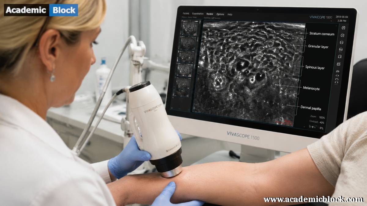
Applications in Dermatology

A. Skin Imaging:

One of the primary applications of RCM is in dermatology, where it has become an invaluable tool for non-invasive skin imaging. RCM allows dermatologists to examine cellular structures, such as epidermal cells, keratinocytes, and melanocytes, in real-time. This capability has revolutionized the diagnosis and monitoring of various skin conditions, including melanoma, basal cell carcinoma, and inflammatory skin disorders.

B. Melanoma Diagnosis:

Reflectance Confocal Microscopy has emerged as a game-changer in the early detection and diagnosis of melanoma, the deadliest form of skin cancer. By providing high-resolution images of melanocytic lesions, RCM aids in differentiating between benign and malignant skin lesions. This not only enhances diagnostic accuracy but also facilitates timely intervention, significantly impacting patient outcomes.

C. Monitoring Treatment Response:

Beyond diagnosis, RCM plays a crucial role in monitoring the response to dermatological treatments. Whether assessing the effectiveness of topical therapies for psoriasis or evaluating the clearance of actinic keratosis after photodynamic therapy, RCM enables clinicians to observe changes at the cellular level. This capability facilitates personalized treatment strategies and optimizes patient care.

III. Advancements in Reflectance Confocal Microscopy

A. Miniaturization and Handheld Devices:

Recent advancements in RCM technology have led to the development of miniaturized and handheld devices. These portable systems offer increased flexibility and accessibility, allowing for point-of-care imaging in diverse clinical settings. Dermatologists can now perform on-site examinations, reducing the need for invasive procedures and improving patient comfort.

B. Artificial Intelligence Integration:

The integration of artificial intelligence (AI) in RCM data analysis has further propelled the capabilities of this imaging technique. Machine learning algorithms can assist in the automated detection of abnormalities, aiding clinicians in quickly and accurately interpreting complex RCM images. This synergy between technology and medicine holds great promise for enhancing diagnostic precision and efficiency.

C. Multimodal Imaging:

Researchers are exploring the potential of combining RCM with other imaging modalities to obtain comprehensive insights into tissue architecture. Multimodal approaches, such as combining RCM with optical coherence tomography (OCT) or multiphoton microscopy, offer complementary information, enabling a more thorough understanding of tissue morphology and pathology.

IV. Mathematical equations behind the Reflectance Confocal Microscopy

The mathematical equations behind Reflectance Confocal Microscopy (RCM) involve principles from optics and signal processing. While a detailed explanation can be quite complex, I’ll provide an overview of the key equations involved in RCM:

Illumination and Reflection:

In RCM, a laser beam is focused onto a specific tissue layer, and the reflected light is collected by a detector. The intensity of the reflected light (I_reflect) can be described using the reflectance coefficient (R) and the incident intensity (I_incident):

I_reflect = R⋅I_incident ;

Here, R is a dimensionless value between 0 and 1, representing the fraction of incident light that is reflected.

Confocal Pinhole:

RCM employs a confocal pinhole to eliminate out-of-focus light, allowing for optical sectioning. The detected signal (Idetected) is given by the convolution of the reflected light intensity with the point spread function (PSF) of the microscope and the transmission function of the confocal pinhole.

Idetected(x,y,z) = ∭R(x′,y′,z′) ⋅ Iincident(x−x′,y−y′,z−z′) ⋅ PSF(x′,y′,z′) dx′ dy′ dz′ ;

Here, (x, y, z) represents the spatial coordinates.

Depth-Resolved Reflectance:

To achieve depth-resolved imaging, the detected signal is often analyzed at different depths (z). The intensity profile along the axial (depth) direction is given by:

Idepth(z) = ∬R(x,y,z) ⋅ Iincident(x,y,z)⋅PSF(x,y,z) dx dy ;

This equation describes how the intensity of the reflected light varies with depth, allowing for the creation of depth-resolved images.

Signal-to-Noise Ratio (SNR):

The SNR is a crucial parameter in microscopy, representing the ratio of the signal intensity to the noise. In RCM, the SNR is influenced by factors such as laser power, detector sensitivity, and the presence of noise sources. Mathematically, SNR is expressed as:

SNR=Signal / Noise ;

High SNR values are desirable for obtaining clear and accurate images.

It’s important to note that the specific implementation and mathematical details may vary based on the exact setup of the RCM system and the algorithms used for image processing and analysis. Researchers and engineers working on RCM continually refine and adapt these equations to improve the performance and capabilities of the technology.

V. Challenges and Future Perspectives

Depth Limitations:

Reflectance Confocal Microscopy is not without its challenges, and one of the primary limitations is the depth of imaging. The penetration depth is restricted, particularly in tissues with high scattering properties. Ongoing research aims to address this limitation through the development of advanced imaging technologies and novel optical techniques.

Clinical Integration:

Despite the significant strides in RCM technology, widespread clinical integration remains a challenge. Overcoming barriers related to cost, training, and standardization is crucial for realizing the full potential of RCM in routine clinical practice. Collaborative efforts between researchers, clinicians, and industry stakeholders are essential to bridge this gap.

Emerging Applications:

As Reflectance Confocal Microscopy continues to evolve, researchers are exploring new avenues and applications. From studying neurological disorders to investigating cellular dynamics in other organ systems, the potential applications of RCM are vast. Ongoing research endeavors are likely to uncover novel uses for this powerful imaging tool in various fields of medicine.

Final Words

In this article by Academic Block we have seen that, the Reflectance Confocal Microscopy stands at the forefront of modern imaging technologies, offering a window into the microscopic world of living tissues. Its applications in dermatology have transformed the diagnosis and management of skin conditions, with far-reaching implications for patient care. As technological advancements continue to push the boundaries of RCM, the future holds exciting possibilities for its integration into diverse medical specialties, promising a deeper understanding of cellular processes and diseases. The journey of Reflectance Confocal Microscopy from the laboratory to the clinic underscores its potential to revolutionize the way we perceive and study the intricacies of life at the cellular level. Please provide your comments below, it will help us in improving this article. Thanks for reading!

This Article will answer your questions like:

+ What is Reflectance Confocal Microscopy (RCM)? >

Reflectance Confocal Microscopy (RCM) is an advanced imaging technique that enables high-resolution, non-invasive visualization of biological tissues at near-cellular level detail. It uses reflected light from tissue structures to create images without the need for tissue removal or staining, making it valuable for dermatological and pathological assessments.

+ How does Reflectance Confocal Microscopy work to image biological tissues? >

RCM works by directing a laser beam onto the tissue surface, where light is reflected back from various depths. A pinhole aperture allows only the reflected light from a specific focal plane to reach the detector, enabling optical sectioning and high-resolution imaging of cellular and subcellular structures within the tissue.

+ What are the key principles and mechanisms of Reflectance Confocal Microscopy? >

RCM utilizes the principle of confocal microscopy combined with reflectance imaging. It employs a laser light source to illuminate the tissue and a pinhole aperture to reject out-of-focus light, allowing only in-focus light to form the image. This enables optical sectioning and depth-resolved imaging of biological structures, providing detailed insights into cellular morphology and tissue architecture.

+ How does RCM compare to traditional light microscopy and histopathology? >

RCM offers advantages over traditional light microscopy and histopathology by providing real-time, non-invasive imaging of living tissue without the need for tissue processing or staining. It enables immediate visualization of cellular structures and dynamic processes in vivo, which is not feasible with conventional microscopy or histopathological techniques that require tissue fixation and sectioning.

+ What types of skin and tissue structures can be visualized using RCM? >

RCM can visualize various skin and tissue structures, including epidermal layers, dermal papillae, hair follicles, and sweat glands. It provides detailed imaging of cellular morphology and subcellular structures such as keratinocytes, melanocytes, and collagen fibers, making it useful for diagnosing skin cancers, inflammatory conditions, and dermatological disorders.

+ What are the advantages of RCM for non-invasive imaging compared to biopsy? >

RCM offers significant advantages over biopsy for non-invasive imaging, including immediate visualization of tissue morphology and cellular details without the need for tissue removal or histological processing. It allows for real-time assessment of skin lesions, reducing patient discomfort and the risk of complications associated with invasive procedures. RCM also facilitates longitudinal monitoring of treatment response and disease progression, providing clinicians with timely diagnostic information and enhancing patient management in dermatology and oncology.

+ How is depth information obtained in Reflectance Confocal Microscopy? >

Depth information in RCM is obtained through optical sectioning, where a pinhole aperture selectively detects light reflected from a specific focal plane within the tissue. By varying the depth of focus using the objective lens, RCM generates sequential images at different depths, enabling three-dimensional reconstruction and visualization of tissue layers and cellular structures.

+ What are the main components of a Reflectance Confocal Microscopy setup? >

A Reflectance Confocal Microscopy setup includes a laser light source (commonly a diode laser), an objective lens with high numerical aperture for focusing and optical sectioning, a pinhole aperture to reject out-of-focus light, and a sensitive photodetector (typically a photomultiplier tube or avalanche photodiode) for capturing reflected light. It also includes scanning mechanisms, optical filters, and image acquisition software for real-time display and analysis of confocal images.

+ How does RCM contribute to the diagnosis of skin cancers and dermatological conditions? >

RCM contributes to the diagnosis of skin cancers and dermatological conditions by providing high-resolution imaging of cellular morphology, aiding in the identification of malignant features such as cell atypia, architectural disarray, and tumor invasion patterns. It enables early detection, differential diagnosis, and assessment of treatment response in conditions like melanoma, basal cell carcinoma, squamous cell carcinoma, and inflammatory skin diseases.

+ What are the limitations and challenges of Reflectance Confocal Microscopy? >

Limitations of RCM include its shallow imaging depth (up to 200-300 micrometers), restricting visualization to superficial tissue layers. Challenges include achieving consistent image quality across diverse skin types and lesion locations, as well as interpreting confocal images for subtle cellular changes that may require histopathological correlation. Technical considerations such as laser safety and image artifact minimization are also critical in clinical practice.

+ How is data from RCM images processed and interpreted? >

Data from RCM images are processed using specialized software for contrast enhancement, image registration, and three-dimensional reconstruction. Image analysis includes quantitative measurements of cell morphology, density, and spatial distribution, aiding in diagnostic assessment and monitoring of disease progression. Interpretation involves correlating confocal findings with clinical history and histopathological information to guide clinical decisions and optimize patient management.

+ What role does fluorescence play in Reflectance Confocal Microscopy? >

Fluorescence in RCM is utilized for contrast enhancement and specific labeling of cellular components or structures within the tissue. Fluorescent dyes or markers can be applied topically or systemically to highlight specific cellular features, facilitating targeted imaging and analysis of molecular interactions. Fluorescence imaging techniques enhance the sensitivity and specificity of RCM for studying biological processes and disease mechanisms at the microscopic level.

+ What recent advancements have been made in Reflectance Confocal Microscopy technology? >

Recent advancements in RCM technology include improved laser sources for higher resolution imaging, advanced scanning techniques for faster image acquisition, and enhanced software algorithms for real-time data processing and analysis. Integration of artificial intelligence and machine learning algorithms enables automated image interpretation and diagnostic support, enhancing the clinical utility of RCM in dermatology and oncology. Miniaturization of RCM devices for handheld use and development of portable systems broaden its application in point-of-care diagnostics and telemedicine.

Hardware and software required for Reflectance Confocal Microscopy

Hardware Components:

  1. Laser Source: A laser source is used to provide the illumination necessary for RCM. Common lasers include diode lasers or solid-state lasers with specific wavelengths suitable for imaging biological tissues.
  2. Confocal Microscope: The core of RCM is a confocal microscope modified for reflectance imaging. This includes optics for focusing the laser onto the specimen and collecting the reflected light. The microscope may have multiple objective lenses for different imaging depths.
  3. Detector: A sensitive detector captures the reflected light from the specimen. Photomultiplier tubes (PMTs) are commonly used detectors in confocal microscopy, providing high sensitivity.
  4. Scanner: A scanning mechanism is essential for obtaining images across the specimen. This can be achieved through a combination of galvanometric mirrors and/or acousto-optic deflectors.
  5. Beam Splitter: A beam splitter separates the incident and reflected light paths, directing the reflected light to the detector.
  6. Pinhole: The confocal pinhole is a critical component that eliminates out-of-focus light, enabling optical sectioning. It can be adjusted to control the thickness of the imaging section.
  7. Z-axis Adjustment: Mechanisms for adjusting the focal plane along the Z-axis allow for imaging at different depths within the specimen.
  8. Control Electronics: Electronics control various components of the system, including the laser power, scanning mechanism, and detector sensitivity.
  9. Computer System: A high-performance computer is necessary for real-time image acquisition, processing, and storage. The computer interfaces with the microscope system and controls the scanning process.

Software Components:

  1. Image Acquisition Software: Specialized software controls the acquisition of images from the RCM system. It manages parameters such as laser power, scanning speed, and image resolution.
  2. Image Processing Software: Post-processing software is used to enhance and analyze acquired images. This may involve deconvolution, contrast adjustment, and other image processing techniques to improve the clarity of cellular structures.
  3. 3D Reconstruction Software: For three-dimensional imaging, software capable of reconstructing image stacks into 3D models is essential. This allows for a more comprehensive understanding of the spatial arrangement of structures within the specimen.
  4. Analysis and Measurement Software: Software tools for quantitative analysis and measurement of features within the images. This may include tools for cell counting, morphological analysis, and other metrics relevant to the specific research or clinical application.
  5. Database and Storage Software: RCM generates large amounts of data. Database and storage software help organize and manage the extensive image datasets efficiently.

List Key Discoveries Where Reflectance Confocal Microscopy is used

Early Detection of Melanoma: RCM has significantly advanced the early detection of melanoma, the deadliest form of skin cancer. By providing high-resolution, real-time imaging of skin lesions, RCM assists in distinguishing between benign and malignant moles, contributing to improved diagnostic accuracy and early intervention.

Diagnosis and Monitoring of Skin Cancer: RCM has revolutionized the diagnosis and monitoring of various skin cancers, including basal cell carcinoma and squamous cell carcinoma. The technology enables clinicians to visualize cellular structures and assess the progression of cancerous lesions over time.

Evaluation of Inflammatory Skin Disorders: Researchers and clinicians have used RCM to study inflammatory skin disorders, such as psoriasis and eczema. The technology allows for detailed examination of cellular changes associated with inflammation, aiding in understanding disease mechanisms and optimizing treatment strategies.

Assessment of Cutaneous Tumors and Lesions: Reflectance Confocal Microscopy has been employed in the assessment of a wide range of cutaneous tumors and lesions. From benign lesions to more complex structures, RCM provides valuable insights into the cellular architecture, facilitating accurate diagnosis and treatment planning.

Monitoring Response to Dermatological Treatments: RCM is utilized to monitor the response of skin conditions to various dermatological treatments, including topical therapies, laser treatments, and photodynamic therapy. Real-time imaging enables clinicians to assess treatment efficacy and adjust therapeutic approaches as needed.

Study of Infectious Skin Diseases: Reflectance Confocal Microscopy has been applied in the study of infectious skin diseases, offering insights into the interaction between pathogens and host tissues. The technology allows for non-invasive observation of cellular responses to infections, aiding in research on antimicrobial therapies.

Understanding Hair and Nail Disorders: RCM has been used to investigate hair and nail disorders, providing detailed images of hair follicles, cuticles, and nail structures. This has advanced our understanding of the pathophysiology of conditions such as alopecia and onychomycosis.

Dermatopathology Research: Reflectance Confocal Microscopy has become a valuable tool in dermatopathology research, allowing pathologists to visualize cellular features in situ. This has implications for studying tissue architecture, enhancing diagnostic accuracy, and contributing to the understanding of disease progression.

Characterization of Skin Aging: Researchers have employed RCM to study the effects of aging on the skin at a microscopic level. This includes the assessment of collagen fibers, elastin content, and changes in cellular morphology, contributing to insights into the aging process and potential interventions.

Exploration of Neurological Disorders: RCM is not limited to dermatology, and researchers are exploring its potential applications in studying neurological disorders. By adapting RCM for imaging neural tissues, scientists aim to uncover cellular details relevant to conditions such as neurodegenerative diseases.

Key figures in Reflectance Confocal Microscopy

Reflectance Confocal Microscopy (RCM) was developed by Dr. Giovanni Pellacani, an Italian dermatologist, and Dr. Milind Rajadhyaksha, an engineer and physicist, during the late 1990s. Their collaboration led to the creation of this powerful imaging technique, which has since become a crucial tool in dermatology for non-invasive skin imaging and diagnosis. While both Pellacani and Rajadhyaksha played key roles in the development of RCM, the term “father” of RCM is sometimes attributed to Dr. Milind Rajadhyaksha due to his expertise in optics and engineering, which contributed significantly to the technology’s advancement.

Facts on Reflectance Confocal Microscopy

Non-invasive Imaging: RCM allows for non-invasive imaging of living tissues, eliminating the need for traditional biopsy procedures. This is particularly advantageous in dermatology, where it has transformed the approach to skin imaging.

Principles of Confocal Microscopy: Reflectance Confocal Microscopy is based on the principles of confocal microscopy, which involves the use of a pinhole to eliminate out-of-focus light. This enables optical sectioning and the generation of high-resolution images at specific depths within the specimen.

Lack of Fluorescent Labels: Unlike traditional confocal microscopy, RCM does not rely on fluorescent labels or dyes. Instead, it utilizes the natural reflectance properties of tissues to generate contrast and visualize cellular structures.

Dermatological Applications: RCM has found widespread applications in dermatology for imaging skin at a cellular level. It has become a valuable tool for the diagnosis and monitoring of skin cancers, inflammatory skin disorders, and various other dermatological conditions.

Early Detection of Skin Cancer: One of the major contributions of RCM is its role in the early detection of skin cancer, particularly melanoma. The technology aids dermatologists in distinguishing between benign and malignant lesions with high precision.

Real-time Imaging: RCM provides real-time imaging, allowing clinicians and researchers to observe dynamic cellular processes and changes as they occur. This feature is crucial for monitoring treatment responses and studying the progression of diseases.

Depth-resolved Imaging: The ability of RCM to achieve depth-resolved imaging is essential for studying different layers of tissues. This capability is valuable in dermatology for assessing structures like epidermis, dermis, and the junction between them.

Multimodal Imaging: Researchers often combine RCM with other imaging modalities, such as optical coherence tomography (OCT) or multiphoton microscopy, to obtain complementary information and enhance the overall understanding of tissue morphology.

Automated Image Analysis: With the integration of artificial intelligence (AI), automated image analysis tools are being developed to assist in the interpretation of RCM images. Machine learning algorithms can aid in the detection of abnormalities and improve diagnostic accuracy.

Clinical Applications Beyond Dermatology: While RCM is widely used in dermatology, its applications are expanding into other medical specialties. Researchers are exploring its potential in studying neurological disorders, gastrointestinal diseases, and various other organ systems.

Portable and Handheld Devices: Recent advancements have led to the development of portable and handheld RCM devices, allowing for greater flexibility and accessibility. These devices are particularly useful for point-of-care examinations in diverse clinical settings.

Academic References on Reflectance Confocal Microscopy

Books:

  1. Rajadhyaksha, M., & González, S. (Eds.). (2018). Confocal Laser Microscopy: Principles and Applications in Medicine, Biology, and the Food Sciences. CRC Press.
  2. Pellacani, G., & Guitera, P. (Eds.). (2018). In Vivo Reflectance Confocal Microscopy for Dermatologic Diseases: A Practical Guide. Springer.
  3. Scope, A., & Dusza, S. W. (Eds.). (2016). Atlas of Reflectance Confocal Microscopy in Dermatology. Springer.
  4. González, S., & Rajadhyaksha, M. (Eds.). (2017). Reflectance Confocal Microscopy of Cutaneous Tumors: An Atlas with Clinical, Dermoscopic and Histological Correlations. Springer.
  5. Farnetani, F., & Pellacani, G. (Eds.). (2018). In Vivo Reflectance Confocal Microscopy of Cutaneous Tumors. Springer.

Journal Articles:

  1. Pellacani, G., Cesinaro, A. M., Seidenari, S., & Magnoni, C. (2005). In vivo confocal scanning laser microscopy of pigmented Spitz nevi: Comparison of in vivo confocal scanning laser microscopy with dermoscopy. Journal of the American Academy of Dermatology, 52(1), 125-133.
  2. Guitera, P., Pellacani, G., Longo, C., Seidenari, S., & Avramidis, M. (2009). In vivo reflectance confocal microscopy enhances secondary evaluation of melanocytic lesions. The Journal of Investigative Dermatology, 129(1), 131-138.
  3. Scope, A., Benvenuto-Andrade, C., Agero, A. L. C., & Marghoob, A. A. (2007). In vivo reflectance confocal microscopy imaging of melanocytic skin lesions: Consensus terminology glossary and illustrative images. Journal of the American Academy of Dermatology, 57(4), 644-658.
  4. Longo, C., Pellacani, G., & Malvehy, J. (2013). Reflectance confocal microscopy for in vivo skin imaging. Cold Spring Harbor Perspectives in Medicine, 3(11), a013860.
  5. Cinotti, E., Perrot, J. L., Labeille, B., et al. (2017). Reflectance confocal microscopy for the diagnosis of vulvar lichen sclerosus. JAMA Dermatology, 153(2), 116-122.
  6. Gambichler, T., Moussa, G., & Sand, M. (2016). Reflectance confocal microscopy in routine practice: Perspectives from dermatologists around the globe. Journal of Dermatological Case Reports, 10(1), 1-7.
  7. Pupelli, G., Longo, C., Veneziano, L., Cesinaro, A. M., Piana, S., & Pellacani, G. (2016). Reflectance confocal microscopy for the in vivo detection of leishmaniasis. JAMA Dermatology, 152(10), 1152-1154.
  8. Scope, A., Benvenuto-Andrade, C., & Scope, A. (2008). Multimodal confocal microscopy: A new paradigm in dermatologic imaging. Dermatologic Clinics, 26(4), 473-484.
  9. Persechino, F., Ciardo, S., Pampena, R., Borsari, S., Longo, C., & Pellacani, G. (2018). In vivo reflectance confocal microscopy of mycosis fungoides with large-cell transformation. Journal of the European Academy of Dermatology and Venereology, 32(11), e423-e424.
0 0 votes
Article Rating
Subscribe
Notify of
guest
0 Comments
Oldest
Newest Most Voted
Inline Feedbacks
View all comments
0
Would love your thoughts, please comment.x
()
x