Exploring Surface Plasmon Resonance (SPR) Imaging

Overview
Surface Plasmon Resonance (SPR) Imaging has emerged as a powerful technique for studying molecular interactions with high sensitivity and real-time monitoring capabilities. This innovative technology has found widespread applications in various fields, ranging from biochemistry to material science. In this article by Academic Block, we will explore the principles, instrumentation, and applications of Surface Plasmon Resonance Imaging, exploring its contributions to advancing our understanding of molecular interactions at the nanoscale.
Understanding Surface Plasmon Resonance
Plasmons: The Basics
To comprehend Surface Plasmon Resonance, one must first grasp the concept of plasmons. Plasmons are collective oscillations of electrons in a metal. When light interacts with a metal surface, it can induce a coherent oscillation of the free electrons, giving rise to plasmon resonances. The frequency at which these oscillations occur is highly sensitive to the properties of the surrounding medium.
SPR Phenomenon
Surface Plasmon Resonance occurs when incident light matches the frequency of plasmon oscillations at the metal-dielectric interface, leading to a dramatic increase in the absorption of light. This resonance condition is highly dependent on the refractive index of the adjacent medium, allowing SPR to serve as a sensitive probe for detecting changes in the immediate vicinity of the metal surface.
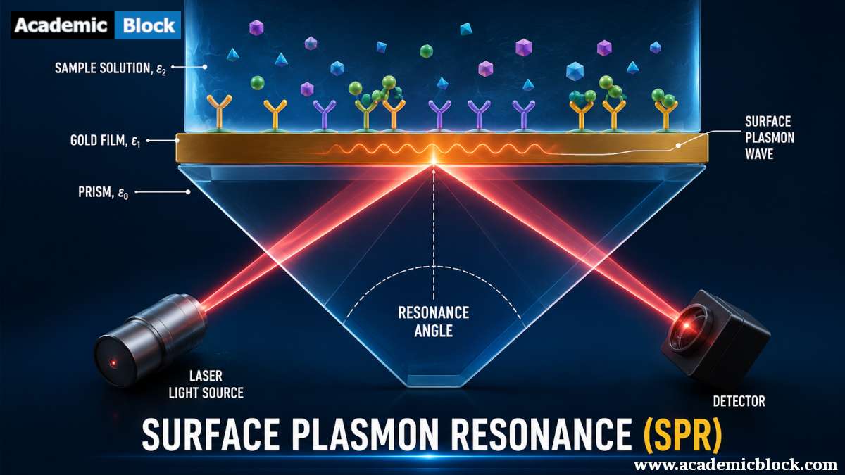
Instrumentation of SPR Imaging
-
Optical Setup: SPR Imaging instruments typically consist of a prism, a light source (commonly a laser), and a detector. The metal film, often gold or silver, is deposited on the prism surface, providing the platform for SPR to occur. The incident light undergoes total internal reflection within the prism, leading to the formation of an evanescent wave at the metal-dielectric interface.
-
Sensor Chip: The sensor chip, where the molecular interactions take place, is crucial for SPR Imaging. It is coated with a thin layer of metal and functionalized with molecules of interest. The binding of analytes to these molecules induces changes in the refractive index at the metal surface, leading to alterations in the SPR signal.
-
Real-Time Monitoring: One of the key advantages of SPR Imaging is its ability to provide real-time data on molecular interactions. As molecules bind or dissociate from the sensor surface, the SPR signal changes, allowing for dynamic monitoring of binding kinetics and affinity.
-
Sensitivity and Resolution: SPR Imaging offers exceptional sensitivity, capable of detecting molecular interactions at the picomolar level. Additionally, the technique provides high spatial resolution, enabling the investigation of interactions at the subcellular and even single-molecule level.
Applications of SPR Imaging
-
Biochemical Research: SPR Imaging has significantly impacted biochemical research by enabling the study of biomolecular interactions in real-time. It is widely employed in the analysis of protein-protein interactions, antigen-antibody binding, and the characterization of enzyme-substrate interactions. The ability to quantify binding kinetics and affinity has enhanced our understanding of various biological processes.
-
Drug Discovery: In the pharmaceutical industry, SPR Imaging plays a crucial role in drug discovery and development. Researchers use this technique to screen potential drug candidates, evaluate their binding affinities to target molecules, and assess the impact of small molecules on protein interactions. The real-time nature of SPR Imaging expedites the drug discovery process by providing rapid feedback on the effectiveness of potential therapeutics.
-
Material Science: Beyond its applications in the life sciences, SPR Imaging has found utility in material science. Researchers use SPR to investigate thin film properties, analyze the adsorption of molecules on surfaces, and study the interactions between materials and biological entities. This versatility has broadened the scope of SPR Imaging, making it a valuable tool in interdisciplinary research.
-
Environmental Monitoring: SPR Imaging is also employed for environmental monitoring and sensing. It can be utilized to detect pollutants, monitor water quality, and study the interactions between environmental samples and specific molecules. The high sensitivity of SPR Imaging makes it an attractive option for applications where precise detection of trace substances is essential.
Mathematical equations behind the Surface Plasmon Resonance Imaging
The mathematical description of Surface Plasmon Resonance (SPR) and Surface Plasmon Resonance Imaging (SPRi) involves several equations that capture the physics of light interacting with a metal surface and the changes in the optical properties due to molecular interactions. Here, we'll outline some key equations associated with SPR and SPRi.
1. Dispersion Relation: The dispersion relation describes the relationship between the wave vector of the incident light and the surface plasmon wave vector. It's given by:
kSP = ( ω / c ) sqrt [ εm / (εm + εd) ] ; where:
- kSP is the surface plasmon wave vector,
- ω is the angular frequency of the incident light,
- c is the speed of light,
- εm is the dielectric constant of the metal,
- εd is the dielectric constant of the dielectric medium.
2. Angle of Resonance: The angle of resonance (θSPR) is the angle at which the surface plasmon resonance occurs. It is given by:
neff(λ) sin(θSPR(λ)) = ( ω / c ) sqrt [ εm / (εm + εd) ] ; where:
-
neff is the effective refractive index of the dielectric medium.
3. Reflectivity Equation: The reflectivity of light at the metal-dielectric interface can be described using the Fresnel equations. For p-polarized light, the reflectivity (R) is given by:
R=∣ (r12 + r23 e2iϕ) / (1 + r12 r23 e2iϕ) ∣2 ;
where:
- r12 is the amplitude reflection coefficient at the metal-dielectric interface,
- r23 is the amplitude reflection coefficient at the dielectric-medium interface,
- ϕ is the phase change upon reflection.
4. Change in Refractive Index: In the context of SPR imaging, the change in refractive index (Δn) due to molecular interactions is a critical parameter. This change is related to the shift in resonance angle (ΔθSPR) by the following equation:
Δn = [ tan(θSPR) / L ] ΔθSPR ; where:
-
L is the penetration depth of the evanescent wave.
5. Binding Kinetics: In SPR imaging, the interaction between molecules on the sensor surface can be characterized using a binding kinetics model, often represented by the Langmuir equation:
θ(t) = θmax [ (ka c) / (1 + kac) ] eZ ; Z = − kdt ;
where:
- θ(t) is the surface coverage at time tt,
- θmax is the maximum surface coverage,
- ka is the association rate constant,
- kd is the dissociation rate constant,
- c is the concentration of the analyte.
These equations provide a mathematical foundation for understanding and analyzing the principles behind SPR and SPR imaging. Researchers use them to interpret experimental data, determine binding kinetics, and optimize experimental conditions for specific applications.
Recent Advances in Surface Plasmon Resonance Imaging
-
Integration with Microfluidics: Recent advancements in SPR Imaging involve the integration of microfluidic systems. Microfluidic devices coupled with SPR Imaging allow for controlled and continuous flow of analytes over the sensor surface, enhancing the efficiency of molecular interaction studies. This integration is particularly valuable for applications requiring minimal sample volumes.
-
Imaging in Complex Matrices: Traditional SPR techniques may face challenges when dealing with complex sample matrices, such as serum or whole blood. However, recent developments in SPR Imaging techniques have addressed these limitations, enabling the analysis of molecular interactions in complex biological samples. This is a significant step forward, as it aligns with the need to study interactions in environments that more closely resemble physiological conditions.
-
Single-Molecule SPR Imaging: Advancements in SPR Imaging have pushed the limits of sensitivity, allowing for the detection and monitoring of single molecules. This breakthrough opens up new avenues for studying molecular interactions at an unprecedented level of detail. Single-molecule SPR Imaging has the potential to unravel intricacies in binding kinetics and uncover nuances that might be masked in ensemble measurements.
Challenges and Future Directions
While SPR Imaging has made remarkable strides, certain challenges persist. The integration of SPR with other imaging modalities, such as fluorescence microscopy, is an area of ongoing research. Combining complementary techniques can provide a more comprehensive understanding of molecular interactions.
Another challenge lies in the development of more robust and versatile sensor surfaces. Improvements in surface functionalization strategies and the exploration of novel materials may further enhance the specificity and stability of SPR sensors.
As technology continues to evolve, the miniaturization of SPR Imaging devices and the development of portable systems are areas of active investigation. These advancements could democratize access to SPR technology, bringing its benefits to a broader range of researchers and industries.
Final Words
Surface Plasmon Resonance Imaging has revolutionized the study of molecular interactions, offering unparalleled sensitivity and real-time monitoring capabilities. From unraveling the intricacies of biochemical processes to advancing drug discovery and material science, SPR Imaging has left an indelible mark on diverse fields.
In this article by Academic Block we have seen that, as the technology continues to advance, the integration of SPR Imaging with other cutting-edge techniques and the exploration of new applications will undoubtedly propel this field further. The ongoing pursuit of higher sensitivity, spatial resolution, and versatility ensures that SPR Imaging will remain at the forefront of molecular interaction studies, pushing the boundaries of our understanding at the nanoscale. Please provide your comments below, it will help us in improving this article. Thanks for reading!
This Article will answer your questions like:
Surface Plasmon Resonance Imaging (SPRi) is a label-free optical technique that measures changes in the refractive index near a sensor surface. It works by detecting surface plasmons, which are oscillations of electrons at the interface between a metal and a dielectric. When biomolecules bind to the sensor surface, they change the local refractive index, which is detected and imaged, allowing for real-time monitoring of molecular interactions.
SPRi is primarily used in biomedical research and diagnostics for analyzing biomolecular interactions, such as protein-protein, protein-DNA, and antibody-antigen interactions. It is also employed in detecting biomarkers for disease diagnosis, studying cellular processes, and evaluating the efficacy and specificity of therapeutic agents.
SPRi detects biomolecular interactions by monitoring changes in the refractive index near the sensor surface. When a target molecule binds to a ligand immobilized on the sensor, it alters the local refractive index. This change is detected by measuring the shift in the resonance angle of surface plasmons, providing real-time data on the interaction kinetics and affinity.
The basic principle of Surface Plasmon Resonance (SPR) involves the excitation of surface plasmons by light at the interface between a metal and a dielectric. When polarized light hits a sensor surface at a specific angle, it induces collective oscillations of free electrons (plasmons), which can be detected as a resonance dip. This shift correlates with changes in the refractive index, indicating molecular binding events.
Surface Plasmon Resonance (SPR) microscopy is primarily used for real-time, label-free monitoring of molecular interactions at surfaces. It is widely employed in biosensing, drug discovery, and material sciences, allowing researchers to study binding events like antigen-antibody interactions or receptor-ligand kinetics without the need for fluorescent or radioactive labels.
Key components of an SPRi system include a light source (usually a laser), a prism or grating to excite surface plasmons, a sensor chip with a metal (typically gold) coating, a detector (such as a CCD camera) to capture the reflected light, and software for data acquisition and analysis. The system also includes fluidics to deliver samples to the sensor surface.
SPRi offers several advantages over ELISA and fluorescence microscopy, including real-time, label-free detection, and the ability to measure binding kinetics directly. Unlike ELISA, which requires labels or secondary antibodies, SPRi can monitor interactions without modifying the biomolecules. Compared to fluorescence microscopy, SPRi provides quantitative data on molecular interactions and is less affected by photobleaching.
SPR is used in a range of applications, including biosensing, drug development, environmental monitoring, and chemical analysis. It enables the study of molecular interactions, such as protein-ligand binding, with real-time data on binding kinetics, specificity, and affinity. SPR's ability to detect interactions without labels makes it ideal for high-throughput screening in pharmaceuticals and biomolecular research.
SPRi's label-free detection avoids potential interference from labels, providing more accurate and reliable data on molecular interactions. It allows for real-time monitoring, capturing dynamic processes and binding kinetics. Additionally, it reduces the time and cost associated with labeling steps, making it suitable for high-throughput screening and analysis.
SPRi is widely used in drug discovery and development for screening potential drug candidates, studying target-ligand interactions, and determining binding affinities and kinetics. It helps identify and characterize lead compounds, optimize drug formulations, and evaluate the specificity and potency of therapeutic agents, accelerating the drug development process.
SPRi plays a crucial role in studying protein-protein interactions by providing real-time, label-free measurements of binding events. It quantifies the association and dissociation rates, allowing researchers to determine binding kinetics and affinities. This information is vital for understanding biological processes, designing inhibitors, and developing therapeutic agents.
Surface Plasmon Resonance Spectroscopy is based on the interaction between light and free electrons in a metal surface, leading to the generation of surface plasmons. By monitoring the angle or wavelength at which resonance occurs, the technique detects changes in the refractive index caused by molecular binding events. This principle is widely used for studying molecular interactions in biosensing and analytical chemistry.
SPRi technology faces limitations such as sensitivity to bulk refractive index changes, limited dynamic range, and potential non-specific binding. Surface regeneration for repeated use and maintaining the stability of immobilized ligands can also be challenging. Additionally, SPRi systems require careful calibration and expertise in data interpretation.
SPR detects changes in the refractive index on a sensor surface, which occur when molecules bind to a thin metal film, typically gold. This allows SPR to detect molecular interactions such as protein-protein, antigen-antibody, and ligand-receptor binding in real-time, without the need for labeling. It can also measure binding affinity and kinetics.
Recent advancements in SPR microscopy include increased sensitivity through the use of nanomaterials and enhanced detection techniques such as localized SPR (LSPR). These advancements have expanded the application of SPR in detecting smaller molecules, providing higher resolution imaging, and enabling the study of single-molecule interactions. Integration with machine learning for data analysis is also becoming more common.
Future developments in SPRi may include integration with other analytical techniques, such as mass spectrometry, for comprehensive biomolecular analysis. Advances in nanotechnology and materials science could lead to more sensitive and specific sensor surfaces. SPRi is also expected to expand in areas like personalized medicine, environmental monitoring, and food safety, providing versatile and powerful tools for various applications.
Hardware and software required for Surface Plasmon Resonance Imaging
Hardware:
- SPR Imaging Instrument: The core of SPRi is the imaging instrument, which includes components like a light source, prism, sensor chip, and optical detection system. Commercial systems from companies like Biacore, Horiba, and GWC Technologies are widely used.
- Sensor Chip: The sensor chip is coated with a thin metal layer (commonly gold) and functionalized with capture molecules. It is crucial for detecting molecular interactions.
- Microfluidic System: In many SPRi setups, a microfluidic system is integrated to control the flow of analytes over the sensor surface. This enhances experimental control and minimizes sample usage.
- Computer: A computer is essential for controlling the SPRi instrument, acquiring data, and running analysis software.
- Temperature Control System: Maintaining a stable temperature is often crucial for reproducibility in SPR experiments. Some SPRi systems include temperature control modules.
- Autosampler: For high-throughput experiments, an autosampler can be integrated to automate sample injections.
Software:
- Control Software: Each SPRi instrument comes with proprietary control software that allows users to set up experiments, control instrument parameters, and monitor real-time data acquisition.
- Data Analysis Software: Analyzing SPRi data involves fitting curves, determining kinetic parameters, and extracting relevant information. Software like Scrubber, TraceDrawer, and BiaEvaluation are commonly used for this purpose.
- Image Analysis Software: For SPRi, capturing images is essential. Image analysis software helps in processing and visualizing the spatial distribution of binding events. Image Studio, GWC Technologies’ SPRimager, and custom MATLAB scripts are examples.
- Statistical Analysis Software: Statistical software like GraphPad Prism or R is often used for in-depth statistical analysis of experimental data and for generating publication-ready graphs.
- Modeling Software: Molecular interaction models are used to simulate and understand experimental outcomes. Software such as MATLAB, Mathematica, or specialized modeling tools can be employed.
- Database Management System: For labs conducting multiple experiments, a database management system can be useful for storing and retrieving experimental data efficiently.
- Surface Plasmon Resonance Instrument Interface Software: Some labs may require software that allows the SPRi instrument to interface with other lab equipment or software systems for broader experimental setups.
- Communication Software: Collaboration tools and communication software facilitate data sharing and discussion among researchers working on SPR experiments.
Key Discoveries made using Surface Plasmon Resonance Imaging
Surface Plasmon Resonance Imaging (SPRi) has been instrumental in advancing our understanding of molecular interactions at the nanoscale. Numerous key discoveries have been made across various scientific disciplines using this powerful technique. Here are some notable discoveries made using SPRi:
- Drug Discovery and Development: SPRi has played a crucial role in drug discovery by providing a high-throughput method for screening potential drug candidates. Researchers can study the binding affinities of small molecules to target proteins, assess the impact of drugs on molecular interactions, and optimize lead compounds for therapeutic development.
- Studying Cellular Processes: SPRi has been used to investigate cellular processes at the molecular level. Researchers have studied cell adhesion, migration, and signaling events by immobilizing cells or cellular components on SPRi sensor surfaces. This has led to insights into the mechanisms underlying various physiological and pathological processes.
- Characterization of Biomaterials and Nanoparticles: SPRi has been employed to study the interaction of biomaterials and nanoparticles with biological molecules. This includes the adsorption of proteins onto surfaces, the binding of nanoparticles to cell receptors, and the development of functionalized surfaces for specific applications in biomedicine and nanotechnology.
- Understanding Enzyme Kinetics: Enzyme-substrate interactions and enzyme kinetics have been investigated using SPRi, providing detailed insights into the catalytic activity of enzymes. This has implications for understanding metabolic pathways, drug metabolism, and the design of enzyme inhibitors.
- Exploration of Microbial Interactions: SPRi has been applied to study interactions involving microorganisms, such as bacteria and viruses. Researchers have used SPRi to investigate microbial adhesion, biofilm formation, and the development of diagnostic assays for infectious diseases.
- Probing Protein Conformational Changes: SPRi has been utilized to study conformational changes in proteins. By immobilizing proteins on sensor surfaces, researchers can monitor changes in protein structure in response to various stimuli, providing valuable information for understanding protein folding and function.
- Single-Molecule SPR Imaging: Advancements in SPRi technology have enabled the detection and monitoring of single molecules. This capability has opened up new possibilities for studying molecular interactions with unprecedented detail, allowing researchers to observe individual binding events and fluctuations in real-time.
- Environmental Monitoring and Sensing: SPRi has found applications in environmental monitoring, including the detection of pollutants and monitoring water quality. The high sensitivity of SPRi makes it suitable for studying interactions between environmental samples and specific molecules, contributing to the development of sensors for environmental analysis.
- Investigating Biophysical Properties of Membranes: SPRi has been employed to study the interactions of molecules with lipid membranes. This has provided insights into the biophysical properties of membranes, including lipid-protein interactions, membrane permeability, and the development of drug delivery systems.
Key Figures in Surface Plasmon Resonance Imaging
The term “Surface Plasmon Resonance” (SPR) and the development of the basic principles can be attributed to Otto Stern and Boris Borisovich Derjaguin, who independently contributed to the theoretical foundations in the 1920s and 1930s.
However, it should be noted that the development of Surface Plasmon Resonance Imaging (SPRi), which involves the imaging and visualization of SPR phenomena, has been a collaborative effort involving contributions from various researchers and scientists. The SPRi technique itself has been developed and refined by multiple researchers in the field of optics, chemistry, and biophysics. Significant contributions have come from scientists like Jiri Homola, Janos Schanda, and many others who have advanced the technology and its applications.
Facts on Surface Plasmon Resonance Imaging
Principle of SPR: SPRi is based on the principle of Surface Plasmon Resonance, which occurs when incident light matches the frequency of plasmon oscillations at the metal-dielectric interface. This resonance condition is highly sensitive to changes in the refractive index near the metal surface, making it an excellent tool for studying molecular interactions.
Real-Time Monitoring: One of the distinctive features of SPRi is its capability for real-time monitoring of molecular interactions. Changes in the refractive index at the metal surface, induced by binding events, can be observed and quantified in real-time.
High Sensitivity: SPRi offers high sensitivity, allowing the detection of molecular interactions at very low concentrations. This makes it a valuable tool for studying biological and chemical interactions with high precision.
Spatial Resolution: SPRi provides spatial information about molecular interactions on the sensor surface. This spatial resolution enables the visualization of binding events at different locations on the sensor chip, offering insights into the distribution of interactions.
Surface Plasmon Resonance Application in Life Sciences: SPRi has been extensively used in the life sciences for studying a wide range of interactions, including protein-protein interactions, antigen-antibody binding, DNA hybridization, and cell-membrane interactions. It has applications in drug discovery, biomolecular kinetics, and understanding cellular processes.
Microarray Technology: SPRi is often integrated with microarray technology, allowing the simultaneous analysis of multiple interactions on a single chip. This high-throughput capability is beneficial for screening large numbers of analytes or studying complex biological samples.
Label-Free Detection: SPRi is a label-free detection technique, eliminating the need for fluorescent or radioactive labels. This feature simplifies experimental procedures, reduces artifacts associated with labeling, and is especially advantageous for studying native biomolecules.
Multimodal Imaging: Some advanced SPRi systems offer multimodal imaging capabilities, combining SPR with other imaging modalities such as fluorescence or surface-enhanced Raman spectroscopy. This integration provides complementary information, enhancing the depth of analysis.
Environmental and Material Science Applications: Beyond the life sciences, SPRi has applications in environmental monitoring and material science. It can be used to study the adsorption of molecules on surfaces, analyze thin films, and investigate interactions between materials and biological entities.
Advancements in Single-Molecule Detection: Recent advancements in SPRi technology have enabled the detection and monitoring of single molecules. This breakthrough has opened new avenues for studying molecular interactions at an unprecedented level of detail.
Commercial Instrumentation: Several companies, such as Biacore, Horiba, GWC Technologies, and others, offer commercial SPRi instrumentation, making the technology accessible to researchers in academia and industry.
Academic References on Surface Plasmon Resonance Imaging
Books:
- Hocker, L. O., & Bechtel, J. M. (Eds.). (2013). Handbook of Surface Plasmon Resonance (Vol. 22). Royal Society of Chemistry.
- Homola, J. (2006). Surface Plasmon Resonance Based Sensors. Springer.
- Liedberg, B., Nylander, C., & Lundström, I. (1983). Surface plasmon resonance for gas detection and biosensing. Sensors and Actuators, 4, 299-304.
- Piliarik, M., & Homola, J. (2009). Surface plasmon resonance (SPR) sensors: approaching their limits? Optics Express, 17(19), 16505-16517.
- Rich, R. L., & Myszka, D. G. (2000). Advances in surface plasmon resonance biosensor analysis. Current Opinion in Biotechnology, 11(1), 54-61.
- Cooper, M. A. (2003). Optical biosensors in drug discovery. Nature Reviews Drug Discovery, 2(8), 515-528.
- Piliarik, M., & Sandoghdar, V. (2014). Direct optical sensing of single unlabelled proteins and super-resolution imaging of their binding sites. Nature Communications, 5, 4495.
- Zeng, Y., Wang, C., Jin, T., & Gu, C. (2019). A review on functionalized gold nanoparticles for biosensing applications. Plasmonics, 14(4), 851-861.
Journal Articles:
- Rich, R. L., & Myszka, D. G. (2001). BIACORE J: A new platform for routine biomolecular interaction analysis. Journal of Molecular Recognition, 14(4), 223-228.
- Homola, J., Yee, S. S., & Gauglitz, G. (1999). Surface plasmon resonance sensors: review. Sensors and Actuators B: Chemical, 54(1-2), 3-15.
- Cooper, M. A., & Singleton, V. T. (2007). A survey of the 2006 literature on optical biosensors. Journal of Fluorescence, 17(4), 329-359.
- Liedberg, B., & Lundström, I. (1993). Principles of biosensing with an extended coupling matrix and surface plasmon resonance. Sensors and Actuators B: Chemical, 11(1-3), 63-72.
- Masson, J. F., & Gao, J. (2013). Direct detection of protein biomarkers in human fluids using site-specific antibody immobilization strategies. Sensors, 13(5), 6045-6057.
- Wang, C., Irudayaraj, J., & Jiang, H. (2010). Activated carbon coated surface plasmon resonance biosensor for pathogen detection. Talanta, 80(1), 313-319.
- Xu, W., Xie, W., Huang, Y., & Wen, Y. (2015). Silver nanoparticles-catalyzed surface plasmon resonance sensing for amplified signal detection of ochratoxin A. Biosensors and Bioelectronics, 68, 213-218.
- Sebba, D. S., Powers, A. S., & Johnson, A. W. (2017). Optical sensor arrays using surface plasmon resonance in conjunction with 3D nanoantenna arrays. Applied Physics Letters, 111(3), 033103.